Near-infrared spectroscopic measurements performed in Japan and a special data analysis method provide new knowledge for a more detailed understanding of certain degenerative processes in plants. The study published by the staff of Kobe University, MATE and ADEXGO Kft. was published in the peer-reviewed journal Plants. The research team has played a significant role in the development and refinement of the aquaphotomics data analysis methodology, and a number of different applications have been reported in recent years.
Water spectral patterns reveals similarities and differences in rice germination and induced degenerated callus development
Zoltan Kovacs, Jelena Muncan, Nobuko Ohmido, George Bazar, Roumiana Tsenkova
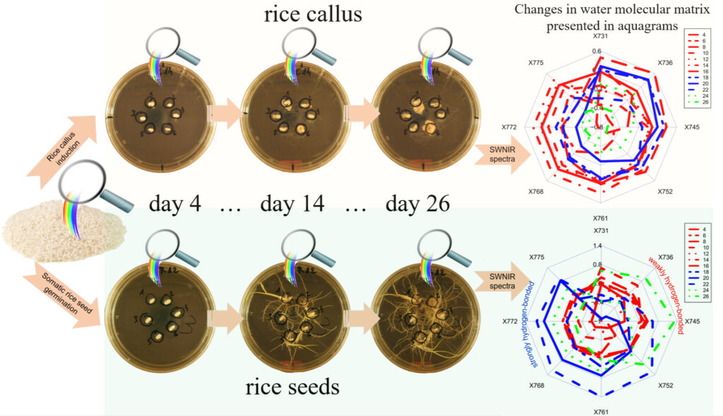
In vivo monitoring of rice (Oryza sativa L.) seed germination and seedling growth under general conditions in closed Petri dishes containing agar base medium at room temperature (temperature = 24.5 ± 1 °C, relative humidity = 76 ± 7% (average ± standard deviation)), and induced degenerated callus formation with plant growth regulator, were performed using short-wavelength near-infrared spectroscopy and aquaphotomics over a period of 26 days. The results of spectral analysis suggest changes in water absorbances due to the production of common metabolites, as well as increases in biomass and the sizes of the samples.
Quantitative models built to predict the day of the development provided better accuracy for rice seedlings growth compared to callus formation. Eight common water bands were identified as presenting prominent changes in the absorbance pattern. The water matrix of only rice seedlings showed three developmental stages: firstly expressing a predominantly weakly hydrogen-bonded state, then a more strongly hydrogen-bonded state, and then, again, a weakly hydrogen-bonded state at the end. In rice callus induction and proliferation, no similar change in water absorbance pattern was observed.
The presented findings indicate the potential of aquaphotomics for the in vivo detection of degeneration in cell development.

Access the full paper free of charge on the website of the journal:
